Aims
SRS treatment planning is based on multimodal imaging, as the co-registration of Magnetic Resonance Imaging (MRI) and CT images, to improve the accuracy of targets definition, but MRI data may contain geometric distortions that could affect precision of co-registration and consequently, the accuracy of Gross Tumor Volume (GTV) delineation.
We analyzed the impact of MRI-related distortion in SRS planning for patients with brain mets.
Materials and Methods
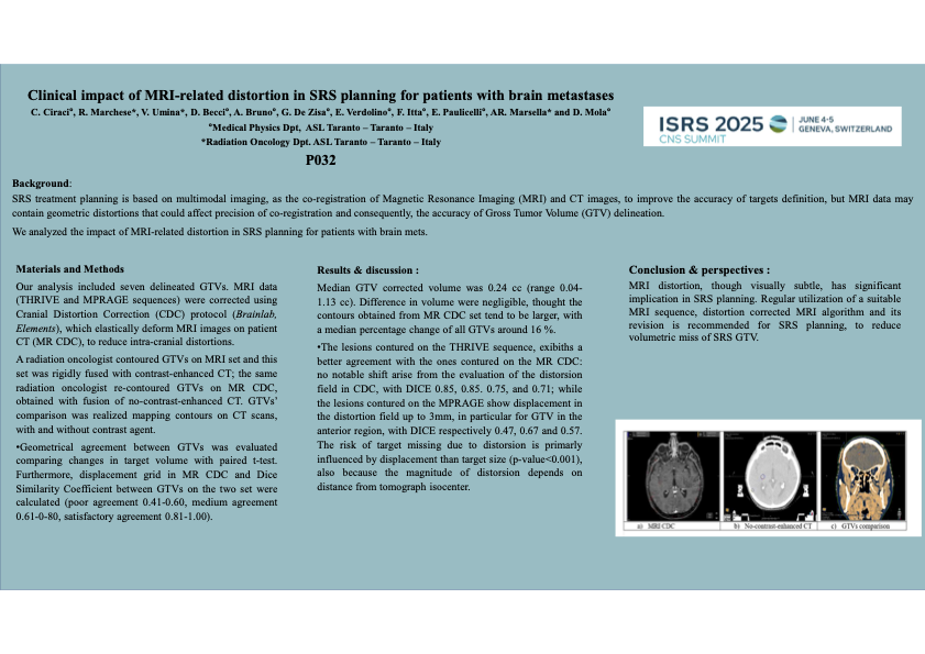
Our analysis included seven delineated GTVs. MRI data (THRIVE and MPRAGE sequences) were corrected using Cranial Distortion Correction (CDC) protocol (Brainlab, Elements), which elastically deform MRI images on patient CT (MR CDC), to reduce intra-cranial distortions.
A radiation oncologist contoured GTVs on MRI set and this set was rigidly fused with contrast-enhanced CT; the same radiation oncologist re-contoured GTVs on MR CDC, obtained with fusion of no-contrast-enhanced CT. GTVs’ comparison was realized mapping contours on CT scans, with and without contrast agent.
Geometrical agreement between GTVs was evaluated comparing changes in target volume with paired t-test. Furthermore, displacement grid in MR CDC and Dice Similarity Coefficient between GTVs on the two set were calculated (poor agreement 0.41-0.60, medium agreement 0.61-0-80, satisfactory agreement 0.81-1.00).
Results
Median GTV corrected volume was 0.24 cc (range 0.04-1.13 cc). Difference in volume were negligible, thought the contours obtained from MR CDC set tend to be larger, with a median percentage change of all GTVs around 16 %.
The lesions contured on the THRIVE sequence, exibiths a better agreement with the ones contured on the MR CDC: no notable shift arise from the evaluation of the distorsion field in CDC, with DICE 0.85, 0.85. 0.75, and 0.71; while the lesions contured on the MPRAGE show displacement in the distortion field up to 3mm, in particular for GTV in the anterior region, with DICE respectively 0.47, 0.67 and 0.57. The risk of target missing due to distorsion is primarly influenced by displacement than target size (p-value<0.001), also because the magnitude of distorsion depends on distance from tomograph isocenter.
Conclusions
MRI distortion, though visually subtle, has significant implication in SRS planning. Regular utilization of a suitable MRI sequence, distortion corrected MRI algorithm and its revision is recommended for SRS planning, to reduce volumetric miss of SRS GTV.