The role of patient monitoring during the dose delivery of any Stereotactic Radiosurgery (SRS) application is of paramount importance and contributes to the overall effectiveness of the treatment. Any potential deviation in the delivered high-dose levels of radiation could lead to poor tumor control and severe complications to the surrounding normal tissue. Therefore, accurate and precise patient positioning on the treatment couch and patient monitoring during the dose delivery of a single fraction or hypo-fractionated SRS applications are mandatory.
In SRS treatments delivered by linear accelerators, the role of image and surface guidance is very important and becomes vital for the safe and efficient implementation of each clinical procedure. The use of advanced Image Guidance Radiation Therapy (IGRT) and Surface Guidance Radiation Therapy (SGRT) techniques increases significantly in recent years being in line with the rapid growth of SRS. IGRT and SGRT systems combine high-end technology with sophisticated software aiming to improve the treatment outcome by minimizing positional uncertainties. Specifically, the most recent integrated IGRT/SGRT systems provide x-ray imaging through embedded Cone Beam Computed Tomography (CBCT) systems or mounted x-ray tubes and corresponding detectors on the linacs. Surface optical imaging with cutting-edge optical cameras, combined with mounted light sources are utilized. The patient’s 3D surface structure and surface thermal pattern are detected using specially designed thermal cameras, acquiring this way the patient’s external thermal surface. However, the more advanced the hardware and software used by these systems are, the higher level of complexity is introduced, and subsequently advanced End-to-End verification is mandatory. This complexity creates the need for a more realistic simulation of the clinical procedure during End-to-End testing.
RTsafe, following the trends of modern SRS, is continuously developing adapted solutions to the most demanding Quality Assurance (QA) needs. Specially designed anthropomorphic head and spine phantoms have been modified and equipped accordingly, to be used for the End-to-End verification of advanced IGRT/ SGRT systems. The combination of anthropomorphic anatomy and bone/soft tissue equivalency that these phantoms offer is a fact that makes them a must-have tool in every radiotherapy department. A tool for verifying the precision of localization and accuracy of dose delivery.
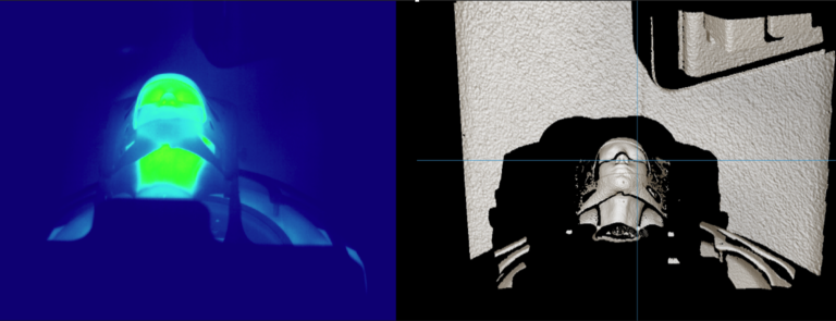
Having the already unique feature of the realistic contrast of bone and soft tissue in CT and MR imaging, the patient-like anatomy, but also the ability to be filled with warm water reproducing human’s external thermal pattern (internal human temperature, heated up to 45°C/113 °F), RTsafe phantoms comprise the best candidate for testing the patient’s positioning and monitoring systems (Figure 1).
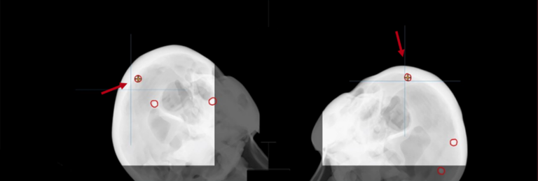
These customized solutions can also accommodate small metal spheres at any desired or predefined location acting as reference points visible in CT, CBCT, MV, and X-ray imaging (Figure 2). The external surface of the phantoms can be modified to simulate realistic external morphological characteristics, with dull surfaces that minimize artifacts and unwanted reflections of optical imaging. The end-users can also perform point and 2D dose measurements using appropriate inserts that fit into the phantoms.
For more information on RTsafe phantoms and services, please visit www.rt-safe.com.